Echocardiogram Testing Clinic in Henderson NV
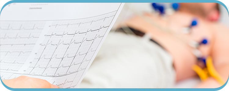
An echocardiogram is a non-invasive ultrasound test that is used to examine the structure and function of the heart. Andrea Warburton MSPHS, PA-C offers echocardiogram tests to identify an irregular heartbeat and other heart conditions, such as heart valve tests, cardiomyopathy, aortic disease, and more. An echocardiogram uses sound waves to create a live image of your heart and it is helpful to determine your heart’s size, blood clot size, surrounding fluid volume, and pressure. To learn more about the testing, contact us or schedule an appointment online. We are conveniently located at 2510 Wigwam Pkwy Suite 102, Henderson, NV 89074.


Table of Content:
What is echocardiogram testing?
What are the different types of echocardiograms?
What conditions require an echocardiogram?
How long does an echocardiogram take?
An echocardiogram is a safe, non-invasive test used to identify and monitor different heart conditions. Detailed images are produced on a computer screen by placing a transducer on the patient’s chest, which emits high-frequency sound waves that bounce off the heart. With these images, medical professionals can assess the structure and operation of the heart. This includes its chamber size and shape, wall thickness and motion, and blood flow through its valves and vessels.
Echocardiograms can also be used to evaluate heart function in patients with known or suspected heart disease, assess heart function during exercise or medication-induced stress tests, or track the development of heart conditions over time. Depending on the type of echocardiogram being performed, the procedure usually lasts 30 to 60 minutes and is painless.
Overall, echocardiogram testing is a useful method for identifying and treating heart disease that assists medical professionals in creating effective treatment plans.
There are several different types of echocardiograms, including:
– Transthoracic echocardiogram (TTE): The most popular type of echocardiogram is a transthoracic echocardiogram, which uses a transducer to produce images of the heart through the chest wall while being applied to the patient’s chest.
– Transesophageal echocardiogram (TEE): To gain closer and more precise images of the heart, this test involves inserting a small, flexible tube with a transducer into the patient’s esophagus.
– Doppler echocardiogram: This type of echocardiogram uses sound waves to measure the speed and direction of blood flow through the heart and blood vessels.
– 3D echocardiogram: This type of echocardiogram uses advanced technology to create a 3D image of the heart, which can provide more detailed information about its structure and function.
– Stress echocardiogram: To assess how the heart responds to stress, this test combines an echocardiogram with physical activity or medication.
– Contrast echocardiogram: This type of echocardiogram uses a contrast dye to make the heart’s structures easier to observe.
– Bubble echocardiogram: Heart structure is tested by injecting a saline solution with microbubbles into a vein. The bubbles travel to the heart and appear on the echocardiogram if a hole is present.
– Fetal echocardiogram: This type is used to evaluate the heart of a developing fetus during pregnancy.
Echocardiograms are used to evaluate and diagnose a wide range of heart conditions, including:
– Heart valve disease: Echocardiograms can identify heart valve abnormalities, such as stenosis, which can cause chest pain, breathlessness, and heart failure.
– Congenital heart disease: Heart defects that are present at birth, such as holes in the heart, that can lead to heart failure and other complications are known as congenital heart disease. Echocardiograms can help identify these defects.
– Cardiomyopathy: Echocardiograms can find heart muscle abnormalities, like thickening or dilation, which can cause heart failure and other complications.
– Pericardial disease: Echocardiograms can identify pericardial disease, which can result in swelling or fluid accumulation around the heart and cause symptoms like chest pain.
– Aortic disease: Echocardiograms can spot aortic abnormalities like aneurysms or dissections, which can be fatal if left untreated.
– Heart failure: Echocardiograms can assess the heart’s ability to pump blood and assess the severity of heart failure, which can help physicians decide what course of action to take.
The type of echocardiogram that is performed and the suspected heart condition will affect the length of the process. For example, a typical transthoracic echocardiogram, the most common type, usually takes between 30 and 60 minutes to complete.
To get the best images, the technician or doctor might have to move the transducer’s position or ask the patient to move. In some circumstances, the technician or doctor may also employ a contrast agent to enhance the visibility of specific heart structures.
The technician or doctor will analyze the images and create a report for the patient’s primary care provider. The results will then be used by the provider to help identify any heart conditions and create a suitable treatment strategy.
To learn more about the testing, contact us or schedule an appointment online. We are conveniently located at 2510 Wigwam Pkwy Suite 102, Henderson, NV 89074. We serve patients from Henderson NV, Midway NV, Gibson Springs NV, Paradise Hills NV, and Winchester NV.


